Achalasia
Achalasia
Esophagus is characterized by its capability to transfer food from mouth to stomach.
This is achieved not only by gravity but also by the peristaltic movements of esophagus in coordination with the relaxation of the sphincter which is placed at the junction of oesophagus and stomach.
Autoimmune derangement of esophageal neurosis results in losing its capability of peristaltic movements and those are replaced by concurrent or absence of peristaltic movements.
This derangement of esophageal neurosis results also to esophageal sphincter inability to relax.
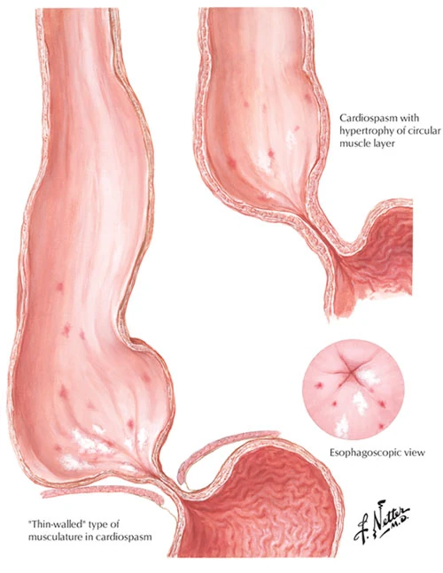
Food stasis gradually distends the esophagus, which is converted to a bag full of food remnants and saliva.
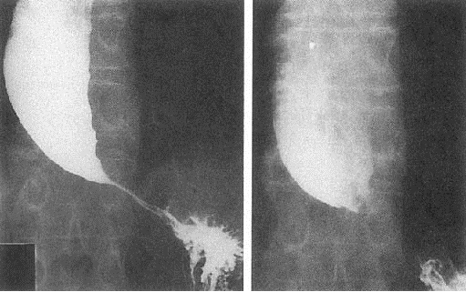
Achalasia diagnosis is based on upper GI endoscopy, esophagogram and manometry.
It is not possible, by any treatment, for the esophagus to regain its normal motility. In the early stages of the disease, swallowing may be improved either by the use of balloon dilatation or by having a cardiomyotomy operation.
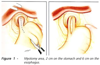
Both forms of treatment aim to reduce the lower esophageal sphincter pressure, so that the food weight will be enough to get it all the way through esophagus to the stomach.

Achalasia-Esophagogram

Cardiomyotomy
Balloon dilatation is preferred for older patients in comparison to cardiomyotomy which is preferred for younger patients or when the dilatation fails.
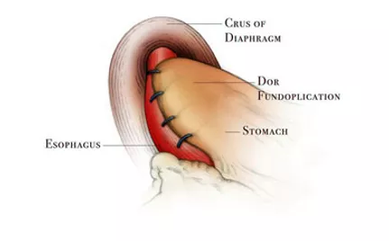
Cardiomyotomy is usually followed by a simple fundoplication, a short of an artificial valve between esophagus and stomach, in order to prevent reflux that is a common consequence of myotomy. Operation success reaches 100% in early stage patients and is reduced to 50% at the last stage of the disease.
Endoscopic cardiomyotomy is performed the last few years in few centers around the world.
Early results are satisfactory but it will take long for it to be used widely since it needs special and long training for the invasive gastroenterologist or the surgeon.
